Annals of Middle Eastern Medicine
Turki Nasser Alotaibi et al. Annals of Middle Eastern Medicine. 2026;2(3):253-258
CASE REPORT
Oral plasmablastic lymphoma in an
HIV-negative immunocompetent patient:
a rare case report
Turki Nasser Alotaibi¹, Ahmed Mufadhi Alanazi²*, Talal Alhumaidi Alshammari³, Sultan Khalid Albaqawi⁴, Mohammed Hassan Aljezoli⁵, Majed Mamdouh Alshammari⁶, Abdulrahman Ibrahim Alsanea⁷, Ebtehal Mohammed Aloudah⁸, Abdulrahman Muteb Alanazi⁸, Alanoud Sultan Alshamari⁹, Esam Sami Almuzaini²
Correspondence to: Ahmed Mufadhi Alanazi
*Department of Periodontology, Hail Dental Center, Hail, Saudi Arabia.
Email: dr.ahmadmufdi@gmail.com
Full list of author information is available at the end of the article.
Received: 06 June 2026 | Revised: 20 June 2026 | Accepted: 22 June 2026
ABSTRACT
Background:
Plasmablastic lymphoma (PBL) is an uncommon and highly aggressive variant of diffuse large B-cell lymphoma, which was first recognized in association with human immunodeficiency virus (HIV). Although an increasing number of cases occurring in HIV-negative immunocompetent individuals have been described, thereby expanding the clinical picture of this tumor.
Methods:
We present a case of a 27-year-old HIV negative, medically fit male who came with a rapidly enlarging gingival mass. Clinical, radiographic, and histopathological assessments were done, plus immunohistochemical staining for plasma cell and B-cell markers. We also checked for Epstein-Barr virus (EBV) and then the Ki-67 proliferation index, basically.
Results:
The patient had about a 2-week history of progressive swelling on the left lower jaw. On examination, there was a lobulated 1.5 × 2.0 cm red to violaceous mass, sitting on the buccal gingiva. Radiographs suggested minimal involvement of the bone. Histopathology showed a diffuse sheet-like infiltrate of atypical plasmablastic cells. In immunohistochemistry, the tumor was positive for CD138, CD38, c-Myc, and EBV, with loss of CD20 expression. The Ki-67 index was above 90%. Altogether, this confirmed the diagnosis of oral PBL.
Conclusion:
Although it is an uncommon entity and usually occurs in patients with HIV infection, oral PBL should be included when comparing treatments for gingival masses that rapidly harden, even in people who do not have AIDS. Patients will have a favorable outcome if they can be identified early, definitely diagnosed by pathologists, and then treated promptly with the proper therapy for this aggressive malignancy.
Keywords:
Plasmablastic lymphoma, oral cavity, HIV-negative, immunocompetent, Epstein-Barr virus, gingival mass.
Introduction
Plasmablastic lymphoma (PBL) is a rare and highly aggressive subtype of diffuse large B-cell lymphoma that was initially described in the oral cavity of patients infected with human immunodeficiency virus (HIV) [1]. Histologically, PBL exhibits plasmablastic morphology and an immunophenotype characterized by expression of plasma cell markers with loss of conventional B-cell markers, particularly CD20 [2,3].
Although PBL is strongly associated with HIV infection and other immunosuppressive conditions, an increasing number of cases have been reported in HIV-negative immunocompetent individuals [4,5]. Oral lesions commonly present as rapidly enlarging gingival or alveolar masses that may mimic benign reactive lesions, resulting in delayed diagnosis [6,7]. Histopathological examination and immunohistochemistry remain essential for establishing the diagnosis, with tumor cells typically expressing plasma cell markers and demonstrating a high Ki-67 proliferation index [2,8].
We report a rare case of oral PBL in an HIV-negative immunocompetent young male and highlight the diagnostic challenges and clinicopathological features of this uncommon malignancy.
Case Presentation
Clinical presentation
A 27-year-old healthy male was referred to the Oral and Maxillofacial Surgery Department in King Khalid Hospital, Hail, Saudi Arabia, with a 2-week history of rapidly increasing swelling of the left lower jaw. No prior medical history or regular medication was recorded in the patient’s file. He did not smoke or drink alcohol. He had no previous HIV infection, immunosuppressive therapy, or organ transplantation. On clinical examination, the patient had no systemic symptoms of fever, night sweats, or weight loss.
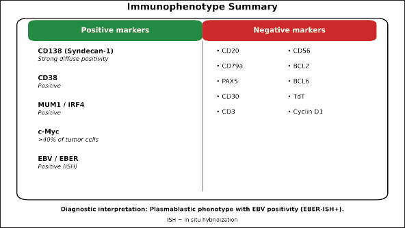
Figure 1. Clinical presentation of the gingival mass. Intraoral photo showing a well circumscribed, lobulated, red-toviolaceous soft tissue mass about 1.5 × 2.0 cm on the buccal gingiva, in relation to tooth #36 (left lower first molar). The lesion seems non-ulcerated with a smoother surface.
Physical examination
Intraorally, a well-circumscribed lobulated soft tissue mass extending from the buccal gingiva in relation to tooth #36 (lower left first molar), measuring 1.5 × 2 cm in its utmost dimension, was noted on examination. The lesion was violaceous-to-red in color, nonulcerative, and smooth-surfaced. The mass was hard on palpation and non-tender. There was no mobility of the associated tooth or pus discharge. The surrounding mucosa appeared normal (Figure 1). Extraoral examination revealed no facial asymmetry or swelling. The cervical lymph nodes were non-palpable. Vital signs of the patient were normal.
Radiographic findings
Panoramic radiograph (orthopantomogram) showed a residual root of tooth #36, but no bone destruction or cortical erosion was depicted. Thus, a peri-apical radiolucency was noted. The radiological appearance was consistent mostly with an extraosseous soft-tissue growth, and there appeared to be no involvement of the bone (Figure 2).
Axial CT scan of the head and neck showed a soft-tissue mass limited to the gingival region without violating the underlying cortical bone. Several enlarged cervical lymph nodes were demonstrated on CT imaging, and lymphoproliferative disease was suspected (Figure 3) Given the clinical presentation and imaging findings, a differential diagnosis was framed, including reactive lesions (pyogenic granuloma and peripheral giant cell granuloma), benign neoplasms, and malignant progressions, including lymphoma.
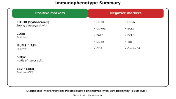
Figure 2. Preoperative panoramic radiograph (orthopantomogram). The radiograph demonstrates a residual root of tooth #36 with associated periapical radiolucency. No evidence of significant bone destruction or cortical erosion is observed, suggesting that the lesion is predominantly extraosseous soft-tissue growth.
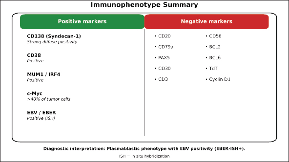
Figure 3. Axial contrast-enhanced CT scan of the head and neck. In this image, there is a soft tissue mass mainly in the gingival area, and it does not show any invasion into the cortical bone below, at all. There are also a few enlarged cervical lymph nodes visible (arrows), and that finding brings up suspicion for a lymphoproliferative disease, or maybe something related - hard to say right away though.
Histopathological findings
The excisional biopsy under local anesthesia discovered microvascular ducts. According to microscopic examination of hematoxylin and eosin-stained sections, normally appearing stratified squamous epithelium covered a diffuse neoplastic infiltrate in the underlying connective tissue. The neoplastic infiltrate consisted of intermediate to large atypical cells arranged in sheets. Tumor cells exhibited eccentric, irregular nuclei with vesicular chromatin, prominent nucleoli, and moderate to abundant basophilic cytoplasm, consistent with the plasmablastic morphology that is commonly found in DLBCL of the plasmablastic variant. Many mitotic figures and apoptotic bodies, as well as tingible body macrophages with nuclei undergoing karyorrhexis or organized clusters, were found in the tumor sections examined under a light microscope. This finding imparts a “starry sky” appearance to plasma cell myxomas and other benign lesions and suggests that it may also be a useful morphologic feature for the diagnosis of plasmablastic tumors in general.
Immunohistochemical analysis
The Ki-67 proliferation index was elevated, estimated at approximately 90%, indicating a highly proliferative neoplasm. The immunophenotypic profile, including expression of plasma cell markers (CD138, CD38, and MUM1) and loss of B-cell markers (CD20, CD79a, and PAX5), c-Myc overexpression with EBV positivity and a very high Ki-67 index, in combination with the plasmablastic morphology, confirmed the diagnosis of a PBL (Figure 4).
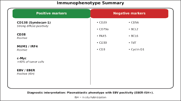
Figure 4. Histopathological and immunohistochemical findings of PBL. (a) Hematoxylin and eosin (H&E) staining shows a diffuse infiltrate of intermediate-to-large atypical cells with eccentric nuclei, vesicular chromatin, prominent nucleoli, and moderate to abundant basophilic cytoplasm, which is consistent with plasmablastic morphology (original magnification ×400). (b) Immunohistochemistry shows strong membranous positivity for CD138, basically a plasma cell marker (original magnification ×400). (c) Strong cytoplasmic expression of CD38 (original magnification ×400). (d) Nuclear positivity for MUM1 (original magnification ×400). (e) Overexpression of c-Myc (original magnification ×400). (f) High proliferative activity, with a Ki-67 index that goes beyond 90% (original magnification ×400). (g) Positive staining for Epstein-Barr virus (EBV) using in situ hybridization (original magnification ×400). (h) Negative staining for CD20, confirming loss of B-cell markers (original magnification ×400).
Table 1. Laboratory findings and staging workup.
| Test/investigation | Finding | Clinical interpretation |
|---|---|---|
| Complete blood count (CBC) | Within normal limits | No cytopenias; no hematologic abnormalities detected |
| Comprehensive metabolic panel (CMP) | Within normal limits | No evidence of hepatic or renal dysfunction |
| Lactate dehydrogenase (LDH) | Elevated | Suggestive of increased cellular turnover and possible tumor burden |
| Ann Arbor stage | Stage IIE | Extranodal involvement (oral cavity) with regional cervical lymph node involvement on the same side of the diaphragm |
Established diagnosis
PBL of the oral cavity (gingival biopsy).
Staging and additional workup
After diagnosis of plasmablast lymphoma, the patient needed a complete initial staging workup (Table 1).
Treatment and follow-up
No therapy was performed in our institution. After the diagnosis was made and initial staging workup was done, the patient was referred to a higher center for further evaluation, multidisciplinary assessment, and definitive treatment planning. The patient was lost to follow-up in our institution after referral, and details of treatment, including the exact chemotherapy regimen and clinical outcomes, are not known.
Discussion
PBL is an uncommon but highly aggressive lymphoma that most frequently involves the oral cavity. Although initially described in association with HIV infection, an increasing number of cases have been reported in HIV-negative immunocompetent patients, expanding the recognized clinical spectrum of the disease [4,5].
The present case demonstrated several characteristic features of oral PBL, including rapid gingival enlargement, minimal radiographic evidence of bone involvement, and histopathological findings consistent with plasmablastic morphology. Because oral PBL may clinically resemble benign reactive lesions such as pyogenic granuloma or peripheral giant cell granuloma, delayed diagnosis is common. Therefore, persistent or rapidly enlarging gingival masses should undergo prompt biopsy and histopathological assessment [6,7].
Accurate diagnosis relies on immunohistochemical evaluation. In our patient, tumor cells expressed plasma cell markers (CD138, CD38, and MUM1) while lacking B-cell markers (CD20, CD79a, and PAX5), together with a Ki-67 proliferation index exceeding 90%. This immunophenotypic profile is characteristic of PBL and assists in distinguishing it from other hematologic and epithelial malignancies [2,3,8].
Epstein-Barr virus (EBV) positivity was identified in the present case. EBV is frequently detected in PBL and is believed to contribute to lymphomagenesis through mechanisms that promote cellular proliferation and survival [9,10]. The presence of EBV in HIV-negative patients further supports its pathogenic role beyond severe immunosuppression.
Despite advances in diagnosis and treatment, PBL remains associated with poor outcomes. Intensive chemotherapy regimens, particularly dose-adjusted EPOCH, are commonly recommended, and recent studies suggest that bortezomib-containing regimens may improve response rates [11]. Because of the rarity of the disease, management generally requires a multidisciplinary approach involving oral and maxillofacial surgeons, pathologists, hematologists, and oncologists.
This case contributes to the growing evidence that oral PBL can occur in young HIV-negative immunocompetent individuals. Early recognition, timely biopsy, and accurate pathological diagnosis remain critical for appropriate referral and management of this aggressive malignancy.
Conclusion
Oral PBL should be considered in the differential diagnosis of rapidly enlarging gingival masses, even in HIV-negative immunocompetent patients. Because its clinical presentation may mimic benign reactive lesions, early biopsy and comprehensive immunohistochemical evaluation are essential for accurate diagnosis. Increased awareness among dental and medical practitioners may facilitate earlier detection, timely referral, and improved management of this rare but highly aggressive lymphoma.
Key learning points
- PBL should be included in the differential diagnosis of rapidly enlarging gingival masses, even in young, HIV-negative, immunocompetent patients without traditional risk factors.
- Accurate diagnosis is the key to an early biopsy and immunohistochemical characterization. PBL usually expresses plasma cell markers (CD138, CD38, and MUM1), lacks B-cell markers (CD20, CD79a, and PAX5), and has a high Ki-67 proliferation index.
- Epstein-Barr virus (EBV) positivity is often associated with PBL and would further support diagnosis in the absence of HIV infection or other forms of immunosuppression.
- Oral PBL may have little bony involvement on radiographs despite rapid clinical growth. Therefore, the absence of radiological destruction should not preclude biopsy of suspicious gingival lesions.
- Early recognition, correct pathological diagnosis, and early referral to a multidisciplinary team (hematologists and oncologists) are essential for the best results in this aggressive malignancy.
Acknowledgments
The authors thank the pathology department at King Khalid Hospital for their assistance with histopathological analysis and immunohistochemistry studies. AI tools were used exclusively for spelling, punctuation, and formatting of Figure 4 only.
List of Abbreviations
CD Cluster of Differentiation
CD20 Cluster of Differentiation 20
CD38 Cluster of Differentiation 38
CD79a Cluster of Differentiation 79a
CD138 Cluster of Differentiation 138 (Syndecan-1)
c-Myc Cellular Myelocytomatosis Oncogene
CT Computed tomography
DLBCL Diffuse Large B-Cell Lymphoma
EBV Epstein–Barr Virus
EPOCH Etoposide, Prednisone, Vincristine (Oncovin), Cyclophosphamide, and Doxorubicin Hydrochloride
HIV Human Immunodeficiency Virus
H&E Hematoxylin and Eosin
IHC Immunohistochemistry
Ki-67 Ki-67 Proliferation Index (Cell Proliferation Marker)
MUM1 Multiple Myeloma Oncogene 1 (IRF4)
OPG Orthopantomogram
PAX5 Paired Box Protein 5
PBL Plasmablastic Lymphoma
Consent to participate
Informed consent was obtained from the patient for publication of this case report and any accompanying images.
Consent for publication
Written informed consent was obtained from the patient for publication of this case report and any accompanying images. A copy of the written consent is available for review by the Editor-in-Chief of this journal.
Availability of data and materials
All data generated or analyzed during this study are included in this published article.
Conflict of interests
The authors declare that they have no competing interests.
Ethics approval
This case report was conducted in accordance with the ethical standards of the institutional research committee of the Ministry of Health, Saudi Arabia
Funding
No funding was received for this case report.
Author details
Turki Nasser Alotaibi¹, Ahmed Mufadhi Alanazi², Talal Alhumaidi Alshammari³, Sultan Khalid Albaqawi⁴, Mohammed Hassan Aljezoli⁵, Majed Mamdouh Alshammari⁶, Abdulrahman Ibrahim Alsanea⁷, Ebtehal Mohammed Aloudah⁸, Abdulrahman Muteb Alanazi⁸, Alanoud Sultan Alshamari⁹, Esam Sami Almuzaini²
- Oral and Maxillofacial Surgery Resident, Hail Dental Center, Hail, Saudi Arabia
- Department of Periodontology, Hail Dental Center, Hail, Saudi Arabia
- Consultant of Oral and Maxillofacial Surgery, King Salman Hospital, Hail, Saudi Arabia
- Oral and Maxillofacial Surgery Specialist, King Khalid Hospital, Hail, Saudi Arabia
- Consultant of Oral and Maxillofacial Surgery, King Khalid Hospital, Hail, Saudi Arabia
- Department of Oral and Maxillofacial Surgery, Maxillofacial Specialist, Ministry of Health, Hail Health Cluster, Hail, Saudi Arabia
- General Dentist, Ministry of Health, Hail Health Cluster, Hail, Saudi Arabia
- Saudi Board Resident (R1), Oral and Maxillofacial Surgery (SB-OMS), Department of Oral and Maxillofacial Surgery, King Khalid Hospital, Saudi Commission for Health Specialties (SCFHS), Hail, Saudi Arabia
- Dental Intern, College of Dentistry, University of Ha’il, Hail, Saudi Arabia
Supplementary content (If any) is available online.
References
- Delecluse HJ, Anagnostopoulos I, Dallenbach F, Hummel M, Marafioti T, Schneider U, et al. Plasmablastic lymphomas of the oral cavity: a new entity associated with the human immunodeficiency virus infection. Blood. 1997;89(4):1413–20. https://doi.org/10.1182/blood.V89.4.1413
- Castillo JJ, Bibas M, Miranda RN. The biology and treatment of plasmablastic lymphoma. Blood. 2015;125(15):2323–30. https://doi.org/10.1182/blood-2014-10-567479
- Campo E, Swerdlow SH, Harris NL, Pileri S, Stein H, Jaffe ES. The 2008 WHO classification of lymphoid neoplasms and beyond: evolving concepts and practical applications. Blood. 2011;117(19):5019–32. https://doi.org/10.1182/blood-2011-01-293050
- Castillo JJ, Winer ES, Stachurski D, Perez K, Jabbour M, Milani C, et al. Clinical and pathological differences between human immunodeficiency virus-positive and human immunodeficiency virus-negative patients with plasmablastic lymphoma. Leuk Lymphoma. 2010;51(11):2047–53. https://doi.org/10.3109/10428194.2010.516040
- Morscio J, Dierickx D, Nijs J, Verhoef G, Bittoun E, Vanoeteren X, et al. Clinicopathologic comparison of plasmablastic lymphoma in HIV-positive, immunocompetent, and posttransplant patients: single-center series of 25 cases and meta-analysis of 277 reported cases. Am J Surg Pathol. 2014;38(7):875–86. https://doi.org/10.1097/PAS.0000000000000234
- Vega F, Chang CC, Medeiros LJ, Udden MM, Cho-Vega JH, Lau CC, et al. Plasmablastic lymphomas and plasmablastic plasma cell myelomas have nearly identical immunophenotypic profiles. Mod Pathol. 2005;18(6):806–15. https://doi.org/10.1038/modpathol.3800355
- Sarode SC, Zarkar GA, Desai RS, Sabane VS, Kulkarni MA. Plasmablastic lymphoma of the oral cavity in an HIV-positive patient: a case report and review of literature. Int J Oral Maxillofac Implants. 2009;38(9):993–9. https://doi.org/10.1016/j.ijom.2009.03.720
- Montes-Moreno S, Martinez-Magunacelaya N, Zecchini-Barrese T, Villambrosía SGD, Linares E, Ranchal T, et al. Plasmablastic lymphoma phenotype is determined by genetic alterations in MYC and PRDM1. Mod Pathol. 2017;30(1):85–94. https://doi.org/10.1038/modpathol.2016.162
- Julia Garcia-Reyero, Nerea Martinez Magunacelaya, Sonia Gonzalez De Villambrosia, Sanam Loghavi, Angela Gomez Mediavilla, Raul Tonda, et al. Genetic lesions in MYC and STAT3 drive oncogenic transcription factor overexpression in plasmablastic lymphoma. Haematologica. 2021;106(4):1120–8. https://doi.org/10.3324/haematol.2020.251579
- Harmon CM, Smith LB. Plasmablastic lymphoma: a review of clinicopathologic features and differential diagnosis. Arch Pathol Lab Med. 2016;140(10):1074–8. https://doi.org/10.5858/arpa.2016-0232-RA
- Castillo JJ, Guerrero‐Garcia T, Baldini F, Tchernonog E, Cartron G, Ninkovic S, et al. Bortezomib plus EPOCH is effective as frontline treatment in patients with plasmablastic lymphoma. Br J Haematol. 2019;184(4):679–82. https://doi.org/10.1111/bjh.15156
Keywords: Plasmablastic lymphoma, oral cavity, HIV-negative, immunocompetent, Epstein-Barr virus, gingival mass.


Publication History
Received: June 06, 2026
Revised: June 20, 2026
Accepted: June 22, 2026
Published: August 15, 2026
Authors
Turki Nasser Alotaibi
Oral and Maxillofacial Surgery Resident, Hail Dental Center, Hail, Saudi Arabia.
Ahmed Mufadhi Marzouq Alanazi
Department of Periodontology, Hail Dental Center, Hail, Saudi Arabia.
Talal Alhumaidi Alshammari
Consultant of Oral and Maxillofacial Surgery, King Salman Hospital, Hail, Saudi Arabia.
Sultan Khalid M Albaqawi
Oral and Maxillofacial Surgery Specialist, King Khalid Hospital, Hail, Saudi Arabia.
Mohammed Hassan Aljezoli
Consultant of Oral and Maxillofacial Surgery, King Khalid Hospital, Hail, Saudi Arabia.
Majed Mamdouh Alshammari
Department of Oral and Maxillofacial Surgery, Maxillofacial Specialist, Ministry of Health, Hail Health Cluster, Hail, Saudi Arabia.
Abdulrahman Ibrahim Alsanea
General Dentist, Ministry of Health, Hail Health Cluster, Hail, Saudi Arabia.
Ebtehal Mohammed Aloudah
Saudi Board Resident (R1), Oral and Maxillofacial Surgery (SB-OMS), Department of Oral and Maxillofacial Surgery, King Khalid Hospital, Saudi Commission for Health Specialties (SCFHS), Hail, Saudi Arabia.
Abdulrahman Muteb Alanazi
Saudi Board Resident (R1), Oral and Maxillofacial Surgery (SB-OMS), Department of Oral and Maxillofacial Surgery, King Khalid Hospital, Saudi Commission for Health Specialties (SCFHS), Hail, Saudi Arabia.
Alanoud Sultan Alshamari
Dental Intern, College of Dentistry, University of Ha’il, Hail, Saudi Arabia.
Esam Sami Almuzaini
Department of Periodontology, Hail Dental Center, Hail, Saudi Arabia.