Annals of Middle Eastern Medicine
Nermeen A. Awad et al. Annals of Middle Eastern Medicine. 2026;2(3):281-289
REVIEW ARTICLE
Cigarette smoking and disease activity, organ damage, and cutaneous manifestations in systemic lupus erythematosus: a systematic review
Nermeen A. Awad1, Lujain Badr Baghlaf2*, Rasha A. Alsulami3, Samah Alotaibi3, Sarah K. Alshehabi3, Wed S. Bin Madhi4
Correspondence to: Lujain Badr Baghlaf
*Bachelor of Medicine, Bachelor of Surgery Program, Fakeeh College for Medical Sciences, Jeddah, Saudi Arabia.
Email: lujainbaghlaf20@gmail.com
Full list of author information is available at the end of the article.
Received: 03 May 2026 | Revised (1): 03 July 2026 |
Revised (2): 07 July 2026 | Accepted: 16 July 2026
ABSTRACT
Background:
Smoking may modify systemic lupus erythematosus (SLE) activity, damage, and phenotype. This review aims to summarize associations between smoking exposure and SLE outcomes.
Methods:
Systematic review of primary studies assessing smoking in SLE and reporting clinical or biomarker outcomes. Five eligible studies (2002-2021) were included (total N = 1,079). Participants were predominantly female (87%-96.2%); where reported, most were Caucasian (73.7%-92%) with mean/median age 37-49 years and disease duration ~9-13.5 years. Smoking exposure definitions varied substantially, limiting direct quantitative pooling and precluding meta-analysis across studies.
Results:
Ever-smoking prevalence ranged ~37%-51%. In one observational study, current smokers had higher disease activity than never smokers [mean total systemic lupus erythematosus disease activity index (SLEDAI) 15.63 ± 1.63 vs. 9.03 ± 0.99; p = 0.0024] and a dose-response by packs/day (p = 0.001). In a retrospective cohort, never exposure to active or second-hand smoke reduced risk of damage accrual (SDI >0: RR 0.78, 95% CI 0.62-0.97; p = 0.0228). Heavy smoking (>20 pack-years) was associated with discoid rash (OR 2.22), photosensitivity (OR 2.19), and neurological disorder (OR 3.16), but inversely associated with renal disorder (OR 0.40), non-erosive arthritis (OR 0.45), and hematological disorder (OR 0.40). Current smoking increased cutaneous damage (any OR 2.73; scarring OR 4.70; active rash OR 6.18). IL-17A levels were similar in SLE and controls, did not correlate with SLEDAI-2K or SDI, and inversely correlated with years of smoking (Rs ≈ -0.43; p ≈ 0.001) Another study reported no significant differences in cumulative damage scores across smoking categories.
Conclusion:
Evidence from five heterogeneous studies links smoking with higher SLE activity and cutaneous damage, and suggests phenotype-specific associations. Standardized exposure measures and prospective designs are needed.
Keywords:
Systemic lupus erythematosus, smoking, autoimmune diseases, risk factors, disease activity.
Introduction
Systemic lupus erythematosus (SLE), often referred to as SLE, is a chronic multisystem autoimmune disease characterized by relapsing inflammation and progressive accrual of irreversible organ damage. Outcomes have improved with earlier recognition and expanding therapeutic options, yet many patients continue to experience flares, disability, and treatment-related toxicity. Long-term complications remain common, and cardiovascular disease is a major driver of morbidity and premature mortality, with meta-analytic evidence indicating that people with SLE have a substantially higher risk of cardiovascular events compared with the general population [1].
Environmental exposures are believed to interact with genetic susceptibility to influence both lupus onset and phenotypic expression. Cigarette smoking remains one of the most prevalent and modifiable exposures relevant to autoimmune disease prevention and management. In large prospective cohort analyses from the Nurses’ Health Study cohorts, smoking was evaluated in relation to incident SLE, and associations differed by anti-double-stranded DNA antibody subtype, supporting biologic heterogeneity in smoking-associated risk [2]. Consistent with this, updated meta-analytic syntheses that adjust for covariates have continued to report that current smoking is associated with higher odds of developing SLE [3].
Several mechanistic pathways provide biologic rationale for smoking-related effects in lupus. Cigarette smoke can increase oxidative stress and endothelial injury, alter innate and adaptive immune responses, and generate modified nucleic acids that may enhance autoantigenicity [4]. Observational work in established SLE has linked smoking with a higher likelihood of anti-double-stranded DNA seropositivity, aligning with pathways that connect nucleic acid sensing to type I interferon signaling [5]. In parallel, contemporary immunology reviews highlight central roles for oxidative stress biology and neutrophil extracellular traps in SLE pathogenesis and tissue injury, processes that can be influenced by reactive oxidant exposure [6].
Clinically, smoking has been repeatedly linked to cutaneous lupus burden and damage. Recent cohort data suggest that higher cumulative exposure measured in pack years is associated with chronic cutaneous manifestations and greater damage accrual, and earlier work reported increased odds of cutaneous damage among current smokers [7]. Smoking has also been associated with reduced responsiveness to antimalarial therapy in cutaneous lupus, creating practical implications for treatment optimization [8]. Reflecting its clinical relevance, European Alliance of Associations for Rheumatology recommendations for SLE management and non-pharmacological care advise assessment of smoking habits and implementation of cessation strategies as part of comprehensive disease management [9].
Despite this mechanistic and epidemiological rationale, the evidence describing how smoking relates to clinical outcomes in patients with established SLE remains limited and fragmented. Most available data derive from single-center studies with modest sample sizes, heterogeneous definitions of smoking exposure, and inconsistent outcome measures, and the reported associations have been variable and at times conflicting across disease activity, organ damage, individual manifestations, and inflammatory biomarkers. No adequately powered multicenter study has been designed primarily to test these associations, and the individual reports have not been formally consolidated, which leaves clinicians without a clear evidence synthesis to guide counselling and risk stratification. A systematic review that brings together evidence from different centers and populations is therefore needed to clarify the direction and consistency of these associations and to define priorities for future research. Accordingly, the objective of this systematic review was to synthesize the available evidence on the association between cigarette smoking exposure and clinical outcomes in SLE, specifically disease activity, cumulative organ damage, individual disease manifestations, cutaneous damage, and inflammatory biomarkers, and to appraise the methodological quality of the underlying studies.
Methods
Protocol registration
This systematic review was conducted in accordance with the Preferred Reporting Items for Systematic Reviews and Meta-Analyses (PRISMA) statement and the Cochrane Handbook for Systematic Reviews [10,11].
Data sources and search strategy
We conducted a comprehensive, filter-free search of PubMed (MEDLINE), Web of Science, Scopus, and the Cochrane Library (CENTRAL) from inception to February 2026. Search terms combined SLE-related keywords and subject headings, when available, with smoking exposure terms. The smoking concept incorporated active smoking and, where applicable, passive exposure terms. Reference lists of included studies and relevant reviews were manually screened to identify additional eligible records. The complete database-specific strategies are provided in Supplementary Table 1.
Eligibility criteria
We included primary human studies evaluating the association between cigarette smoking exposure and outcomes in SLE. Eligible designs were observational studies (cross-sectional analyses, cohort studies, including retrospective cohorts, and other non-randomized analytic designs). Studies were required to include participants with SLE defined using established classification criteria (e.g., American College of Rheumatology criteria and/or SLICC criteria [12,13]), quantify smoking exposure (e.g., pack-years, packs/day, years of smoking, current/former/never, ever/never, or passive exposure), and report at least one pre-specified outcome domain. Outcomes of interest included disease activity systemic lupus erythematosus disease activity index (e.g., SLEDAI or SLEDAI-2K), cumulative organ damage (e.g., SLICC/ACR Damage Index), specific SLE manifestations, cutaneous damage items, and inflammatory biomarkers.
We excluded case reports/series without analytic comparisons, conference abstracts without full data, reviews, editorials, letters, clinical guidelines, book chapters, non-human or in-vitro studies, and articles with overlapping or duplicate datasets, in which case the most complete or most recent publication was retained. Non-English articles were excluded.
Study selection
Search results from all databases were imported into Rayyan (Rayyan Systems Inc., Cambridge, MA) [14] and duplicates were removed. Two reviewers independently screened titles and abstracts, followed by full-text assessment of potentially eligible articles. Disagreements were resolved by discussion and, when needed, adjudication by a third reviewer. Reasons for exclusion at the full-text stage were documented and summarized in the PRISMA flow diagram.
Data extraction
Data extraction was performed independently by two reviewers using a piloted standardized spreadsheet. Extracted variables included study design, setting/country, sample size, participant demographics (age, sex distribution, and ethnicity where reported), disease duration, smoking exposure definition and ascertainment method (including categorization thresholds and capture of second-hand exposure), outcome definitions and instruments (e.g., SLEDAI, SDI, ACR manifestations, cutaneous SDI items), follow-up duration for longitudinal cohorts, and confounders included in adjusted analyses.
We extracted effect estimates as reported (e.g., odds ratios, risk ratios, regression coefficients, mean differences, and correlation coefficients), along with 95% confidence intervals and p-values. When multiple models were presented, we prioritized the most appropriately adjusted model (typically including at least age and sex, and additionally disease duration and treatment where available). Any discrepancies were resolved by consensus, with arbitration by a third reviewer when necessary.
Risk of bias
Risk of bias was independently assessed by two reviewers using the Newcastle-Ottawa Scale adapted for observational studies [15]. Cohort studies were evaluated across selection, comparability, and outcome domains, while cross-sectional studies were assessed using the modified Newcastle-Ottawa Scale appropriate for cross-sectional designs. Disagreements between reviewers were resolved through discussion with a third senior reviewer. Studies were categorized as good, moderate, or low quality according to predefined Newcastle-Ottawa thresholds [16].
Synthesis approach
Given anticipated clinical and methodological heterogeneity in both smoking exposure measurement as pack-years, packs/day, years smoked, and categorical definitions, and outcome ascertainment across studies, we conducted a qualitative synthesis without meta-analysis. Findings were summarized by pre-specified outcome domains including disease activity, cumulative damage, specific manifestations, cutaneous damage, and biomarkers. For each domain, we reported the direction and magnitude of associations, emphasized adjusted estimates when available, and highlighted dose-response patterns and consistency across studies.
Results
Literature search
The PRISMA flow diagram shows that 2,318 records were identified from databases (PubMed/MEDLINE 1,137, Web of Science 680, Scopus 450, and Cochrane CENTRAL 51), with 1,043 duplicate records removed, leaving 1,275 records for screening. After title and abstract screening, 1,245 records were excluded, and 30 reports were sought for retrieval, of which 5 were not retrieved. Twenty-five full-text reports were assessed for eligibility, and 20 were excluded, mainly for not being primary studies or lacking relevant smoking exposure. Ultimately, five studies met the eligibility criteria and were included in the review [17-21] (Figure 1).
Risk of bias assessment
The risk of bias was assessed using the Newcastle-Ottawa Scale. Overall, studies ranged from moderate to good quality (Table 1). Cohort studies demonstrated higher quality, while cross-sectional studies had more bias due to confounding and variability in smoking exposure definitions.
Study selection and characteristics
A total of five studies met the inclusion criteria and were included in this systematic review [17-21]. The included studies were published between 2002 and 2021 and comprised a combined sample of 1,079 SLE patients. Study designs included three cross-sectional analyses, one observational study, and one retrospective cohort study. Individual sample sizes ranged from 105 to 485 participants. All studies enrolled predominantly female populations (87%-96.2%), consistent with the known sex distribution of SLE. Where reported, most participants were Caucasian (73.7%-92%). Mean or median ages at enrolment ranged from approximately 37 to 49 years, and disease duration ranged from approximately 9 to 13.5 years. The characteristics of each included study are summarized in Table 2.
Smoking exposure was measured heterogeneously across studies. Two studies quantified exposure using pack-years, one used pack per day in a dose-response analysis, one used year of smoking as a continuous variable, and one dichotomized exposure as ever versus current smoker status. Montes et al. [19] additionally captured second-hand smoke exposure. The prevalence of ever-smoking among included cohorts ranged from approximately 37% to 51%.
Smoking and SLE disease activity
Two studies investigated the relationship between smoking and disease activity in SLE, measured using different instruments and approaches. Ghaussy et al. [21] directly compared SLEDAI scores across smoking categories and observed significantly higher mean total SLEDAI scores in current smokers (15.63 ± 1.63) compared with ex-smokers (11.46 ± 2.12) and never smokers (9.03 ± 0.99; p = 0.0024). This difference was observed in both the neurological component of the SLEDAI (neuro-SLEDAI: 1.25 ± 0.32 vs. 0.50 ± 0.16; p = 0.003) and the non-neurological component (14.38 ± 1.55 vs. 8.53 ± 0.94; p = 0.003). A significant dose-response relationship was identified, whereby increasing packs per day among current smokers was associated with higher SLEDAI scores (p = 0.001).
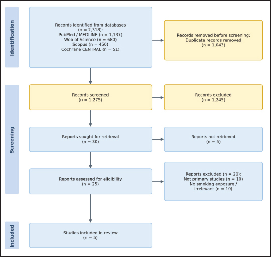
Figure 1. PRISMA flow diagram of study selection.
Table 1. Risk of bias assessment using the Newcastle-Ottawa Scale.
| Study | Selection (max 4) | Comparability (max 2) | Outcome/exposure (max 3) | Total | Quality |
|---|---|---|---|---|---|
| Leffers et al. [20] | 3/4 | 2/2 | 2/3 | 7/9 | Good |
| Ghaussy et al. [21] | 2/4 | 1/2 | 2/3 | 5/9 | Moderate |
| Montes et al. [19] | 3/4 | 2/2 | 3/3 | 8/9 | Good |
| Raymond et al. [17] | 2/4 | 1/2 | 2/3 | 5/9 | Moderate |
| Turchin et al. [18] | 2/4 | 2/2 | 2/3 | 6/9 | Moderate |
NOS, Newcastle-Ottawa Scale. Each domain is scored as the number of points awarded out of the maximum for that domain (selection, 4; comparability, 2; outcome or exposure, 3); the maximum total score is nine points. Item-level justifications for each study are provided in Supplementary Table 2.
Table 2. Characteristics of included studies.
| Author, year | Study design | N | Population | Smoking measure | Key outcomes assessed |
|---|---|---|---|---|---|
| Leffers et al. (2021) [20] | Cross-sectional | 485 | Danish SLE cohort; 88% female; 92% Caucasian; median age 49 years | Pack-years (0, >0, >10, >20); ever/never smoker | ACR-defined SLE manifestations; phenotypic clustering |
| Ghaussy et al. (2002) [21] | Observational | 111 | SLE patients; current (n = 29), ex- (n = 17), never smokers (n = 65); mean age 37-44 years | Current/ex-/never smoker; packs/day (dose-response) | SLEDAI (total, neuro, non-neuro); SLICC/ACR damage |
| Montes et al. (2016) [19] | Retrospective cohort | 105 | SLE patients; 96.2% female; mean age 40.7 years; mean follow-up 9.0 years | Active/secondhand/ current/former smoking; ever exposed versus never | SLICC/ACR Damage Index (SDI) progression to SDI > 0 |
| Raymond et al. (2017) [17] | Cross-sectional | 102 | SLE patients (87% female; mean age 49 years; median SLEDAI-2K 6) and 31 healthy controls | Years of smoking (continuous) | Serum IL-17A levels; correlation with SLEDAI-2K and SDI |
| Turchin et al. (2009) [18] | Cross-sectional | 276 | SLE patients; 92% women; 73.7% Caucasian; mean age 45.1 years; mean disease duration 13.5 years | Ever/current smoker status | Cutaneous damage items from SDI (scarring, alopecia, active rash) |
ACR, American College of Rheumatology; PY, pack-years; SDI, SLICC/ACR Damage Index; SLEDAI, Systemic Lupus Erythematosus Disease Activity Index; y, years.
Raymond et al. [17] also assessed associations between smoking and disease activity (SLEDAI-2K) but approached this through the lens of IL-17A, an inflammatory cytokine. In their cohort, median SLEDAI-2K was 6, and IL-17A levels did not significantly correlate with SLEDAI-2K scores. However, a notable inverse correlation was observed between IL-17A and cumulative years of smoking (Rs ≈ -0.43; p ≈ 0.001), suggesting that prolonged smoking exposure may modulate inflammatory cytokine profiles in SLE patients.
Smoking and organ damage
Two studies specifically examined the relationship between smoking and cumulative organ damage as quantified by the SLICC/ACR Damage Index (SDI). Montes et al. [19] assessed damage progression in a retrospective cohort of 105 SLE patients and found that never-exposed status (neither active nor second-hand smoke) was associated with a significantly reduced risk of any organ damage (SDI >0), with a relative risk of 0.78 (95% CI 0.62-0.97; p = 0.0228), corresponding to an approximate 22% relative risk reduction. Mean SDI scores were numerically lower in unexposed patients (1.80 vs. 2.15), and specific damage items such as scarring alopecia were less frequent among non-exposed individuals (3.1% vs. 10.3%). The accompanying systematic review by the same authors identified only six articles evaluating smoking and SDI, none of which were primarily designed to test this relationship, highlighting the paucity of dedicated research in this area.
In contrast, Ghaussy et al. [21] reported no statistically significant differences in SLICC/ACR damage scores across smoking groups (current, ex-, and never smokers), although a non-significant trend toward higher damage scores in current smokers was noted. The relatively small sample size of current smokers (n = 29) may have limited statistical power to detect such differences.
Smoking and specific SLE manifestations
Leffers et al. [20] conducted the most comprehensive analysis of the association between cumulative smoking exposure (pack-years) and individual SLE manifestations defined by the American College of Rheumatology criteria. Using logistic regression adjusted for age and sex, the authors identified dose-dependent positive associations for several manifestations at the highest exposure stratum (>20 pack-years vs. never smokers). Discoid rash (OR 2.22; 95% CI 1.00-4.92), photosensitivity (OR 2.19; 95% CI 1.13-4.27), and neurological disorder (OR 3.16; 95% CI 1.26-7.96) were all significantly more prevalent among heavy smokers. The association with neurological disorder was particularly robust, persisting after additional adjustment for photosensitivity and discoid rash (OR 3.09; p = 0.018).
Conversely, several manifestations exhibited significant inverse dose-dependent associations with heavy smoking. Renal disorder (OR 0.40; 95% CI 0.19-0.83), non-erosive arthritis (OR 0.45; 95% CI 0.26-0.78), and hematological disorder (OR 0.40; 95% CI 0.20-0.77) were all significantly less prevalent in the >20 pack-year group compared with never smokers. Hierarchical cluster analysis revealed three phenotypic clusters with different smoking prevalence distributions (p = 0.0024), suggesting that smoking may influence the overall phenotypic expression of SLE.
Smoking and cutaneous damage
The relationship between smoking and cutaneous damage was addressed by two studies. Turchin et al. [18] focused specifically on cutaneous items from the SDI in 276 SLE patients and found that current smokers had significantly higher adjusted odds of any cutaneous damage (OR 2.73; 95% CI 1.10-6.81), scarring (OR 4.70; 95% CI 1.04-21.2), and active rash (OR 6.18; 95% CI 1.63-23.3) compared with non-smokers. These findings are consistent with the observation by Leffers et al. [20] that discoid rash and photosensitivity were more common among heavy smokers, as well as the numerical trend reported by Montes et al. [19] showing higher rates of scarring alopecia among smoking-exposed individuals (10.3% vs. 3.1%). These data suggest a convergent pattern in which smoking disproportionately affects the skin in SLE patients.
Table 3. Summary of key findings on smoking and SLE outcomes.
| Author, year | Outcome domain | Key findings | Effect size (95% CI) | p -value |
|---|---|---|---|---|
| Leffers et al. (2021) [20] | SLE manifestations (>20 PY vs. never) | Discoid rash | OR 2.22 (1.00-4.92) | NR |
| Photosensitivity | OR 2.19 (1.13-4.27) | NR | ||
| Neurological disorder | OR 3.16 (1.26-7.96) | 0.018* | ||
| Renal disorder (inverse) | OR 0.40 (0.19-0.83) | NR | ||
| Non-erosive arthritis (inverse) | OR 0.45 (0.26-0.78) | NR | ||
| Hematological disorder (inverse) | OR 0.40 (0.20-0.77) | NR | ||
| Ghaussy et al. (2002) [21] | Disease activity | Total SLEDAI: current 15.63 versus never 9.03 | NR | 0.0024 |
| Neuro-SLEDAI: current 1.25 versus never 0.50 | NR | 0.003 | ||
| Dose–response: ↑ packs/day → ↑ SLEDAI | NR | 0.001 | ||
| Montes et al. (2016) [19] | Organ damage (SDI) | Never-exposed: reduced risk of SDI > 0 | RR 0.78 (0.62-0.97) | 0.0228 |
| Raymond et al. (2017) [17] | Biomarker (IL-17A) | IL-17A inversely correlated with years of smoking | Rs ≈ -0.43 | ≈0.001 |
| Turchin et al. (2009) [18] | Cutaneous damage | Any cutaneous damage | OR 2.73 (1.10-6.81) | NR |
| Scarring | OR 4.70 (1.04-21.2) | NR | ||
| Active rash | OR 6.18 (1.63-23.3) | NR |
CI, confidence interval; IL-17A, interleukin-17A; OR, odds ratio; PY, pack-years; RR, relative risk; Rs, Spearman rank correlation coefficient; SDI, SLICC/ACR Damage Index; SLEDAI, Systemic Lupus Erythematosus Disease Activity Index. *After additional adjustment for photosensitivity and discoid rash. NR, not reported or not applicable.
Smoking and inflammatory biomarkers
Raymond et al. [17] investigated serum IL-17A levels in 102 SLE patients and 31 healthy controls. Median IL-17A concentrations were similar between SLE patients and controls (28.4 vs. 28.4 pg/ml; p ≈ 0.90). Within the SLE cohort, IL-17A did not correlate with disease activity (SLEDAI-2K) or overall cumulative damage (SDI). However, years of smoking was significantly and inversely associated with IL-17A levels (Rs ≈ -0.43; p ≈ 0.001). The authors proposed that IL-17A may play a dual role in SLE pathogenesis, participating in inflammation while potentially exerting a protective function, and that chronic smoking exposure may attenuate this cytokine pathway. The key findings from all included studies, including effect sizes and corresponding statistical significance, are presented in Table 3.
Discussion
Across the five included studies, we found converging signals that smoking exposure is linked to a more severe SLE phenotype, but with heterogeneity by outcome domain and by how smoking was measured. The most consistent pattern in our dataset was the enrichment of cutaneous involvement and cutaneous damage among smokers, alongside higher disease activity in current smokers in the single study that compared SLEDAI across smoking strata.
Our results align with larger epidemiologic work suggesting that current smoking is associated with SLE risk, particularly for anti-double-stranded DNA positive disease, and that smoking is a modifiable exposure that likely interacts with SLE biology rather than acting as a uniform trigger across all subtypes. Large prospective cohort analyses have reported higher risk estimates for anti-double-stranded DNA-positive SLE among current smokers, with weaker or absent associations for overall SLE, when serology is not considered [2]. Meta-analytic syntheses similarly support increased odds of SLE among current smokers versus never smokers, while former smokers often show attenuated risk, suggesting that duration, intensity, and recency of exposure matter [22].
The mixed directionality seen in Leffers et al. [20] with increased odds of some manifestations and lower odds of renal, hematological, and arthritis outcomes at high pack years, contrasts with the general clinical intuition that smoking worsens systemic disease burden [20]. We interpret these inverse associations cautiously as they can arise from selection effects, survival bias, differential treatment patterns, and unmeasured confounding, especially in cross-sectional designs where exposure and outcome are assessed contemporaneously. This is important because longitudinal cohorts outside our inclusion set show that current or past smoking is associated with earlier onset of damage and faster damage accrual [23,24].
Several mechanistic pathways link cigarette smoke to immunologic and end-organ outcomes relevant to SLE. Cigarette smoke contains oxidants and reactive aldehydes that amplify oxidative stress, modify self-antigens, and increase immunogenic cell death products, processes that can feed autoantibody generation and immune complex formation [25]. Reviews focused on smoking and SLE outline effects on B cells and T cells, cytokine networks, oxidative stress biology, and DNA adduct formation that can contribute to loss of tolerance [4].
A second, tightly connected axis is innate immune activation through neutrophil extracellular traps and type I interferon programs. SLE is characterized in many patients by an interferon-regulated gene signature that correlates with activity and specific clinical subsets, and interferon pathway biology is now central to therapeutic development and stratification [26-28]. Neutrophil Extracellular Traps (NET) formation and impaired NET clearance can amplify nucleic acid sensing, activate plasmacytoid dendritic cells, and reinforce interferon production, forming a feed-forward loop that can sustain disease [29,30]. In parallel, experimental and clinical literature indicates that cigarette smoke exposure can promote NET biology in other inflammatory contexts and that nicotine and smoke-related stimuli can induce NET formation, offering a biologic bridge between smoking and lupus-relevant innate immune activation [31,32].
Our included biomarker study suggested an inverse relationship between years of smoking and IL-17A, while showing no clear correlation of IL-17A with activity or damage. Interpreting this requires nuance because IL-17 and Th17 programs are frequently elevated in SLE and are implicated in tissue inflammation, but cytokine levels in serum can be decoupled from tissue-level activity, therapy exposure, and chronic immune exhaustion [33]. Smoking may also exert bidirectional immunomodulatory effects, for example attenuating selected cytokines or altering dendritic cell outputs while still promoting pathogenic autoimmunity through oxidative and epigenetic mechanisms. Human plasmacytoid dendritic cell studies demonstrate that cigarette smoke can alter cytokine production in response to nucleic acid-sensing pathways, supporting the idea that chronic smoke exposure can reshape innate signalling rather than uniformly amplifying it [34].
The strongest coherence between our review and external literature lies in skin disease. Our included Turchin et al. [18] analysis found higher odds of cutaneous damage items among current smokers, and Leffers et al. [20] identified higher odds of discoid rash and photosensitivity at high pack years. In the broader cutaneous lupus literature, active smoking is repeatedly associated with worse disease course, lower likelihood of long-term remission, and reduced responsiveness to antimalarials in many cohorts [35-37]. Mechanistically, proposed explanations include higher baseline disease severity in smokers, adherence differences, altered drug handling, and cellular effects of smoke on lysosomal biology and inflammatory recruitment within the skin [38-40]. This is clinically relevant because hydroxychloroquine is a cornerstone therapy across SLE and cutaneous lupus, and blood level studies show that lower hydroxychloroquine concentrations are associated with higher activity and higher flare risk, implying that any exposure that reduces effective drug exposure or response could translate into worse outcomes [41,42].
Our included studies showed mixed findings for cumulative damage, with one cohort suggesting reduced risk of SDI progression in never-exposed individuals and another finding no clear difference by smoking strata. Outside our dataset, incident SLE cohorts report faster damage accrual and worse composite outcomes among current or former smokers, supporting the interpretation that smoking contributes to irreversible morbidity over time [23,24]. For kidneys, the inverse association with renal disorder at high pack years seen in one cross-sectional study should not be treated as protective. Classical lupus nephritis cohort data indicate that smoking at nephritis onset is associated with faster progression to end-stage renal disease independent of hypertension and immunosuppression, and gene-environment interaction data suggest smoking may potentiate nephritis risk in genetically susceptible patients, such as carriers of STAT4 risk variants [43,44].
Finally, smoking is a major driver of cardiovascular risk in the general population, and cardiovascular disease is a leading contributor to long-term morbidity and mortality in SLE. A systematic review and meta-analysis focused on SLE populations reports higher cardiovascular disease risk among current smokers, reinforcing the need to integrate smoking cessation into cardiovascular risk modification in lupus care [45].
Limitations
Our evidence base is small and dominated by cross-sectional designs, which limits causal inference and makes reverse causation possible, for example, disease severity influencing smoking behavior or cessation. Smoking exposure definitions varied substantially across studies, ranging from ever current status to pack years, years smoked, or second-hand exposure, creating measurement non-equivalence and limiting synthesis. Residual confounding is likely, including socioeconomic factors, medication exposure, adherence, comorbidity burden, and sun exposure patterns for cutaneous outcomes. Only five studies met our eligibility criteria, and the substantial heterogeneity in exposure definitions and outcome measures precluded a quantitative meta-analysis, so the synthesis remained narrative. Restricting eligibility to English-language publications may also have introduced language bias through the omission of relevant non-English reports.
Future directions
Prospective longitudinal cohorts with standardized smoking exposure capture are needed, including time-updated current versus former status, pack years, age at initiation, cessation timing, and second-hand exposure, ideally supplemented by biomarkers such as cotinine. Mechanistic studies should connect smoking exposure to interferon pathway activation, NET biology, and epigenetic remodeling [46-48] using multiomics in well-phenotyped cohorts and with attention to treatment effects. Interventional work should evaluate whether structured cessation programs reduce flare frequency, improve cutaneous outcomes, slow damage accrual, and enhance response to hydroxychloroquine or other therapies.
Implications for practice
Given the consistency of associations with cutaneous disease and the accumulating longitudinal evidence linking smoking to earlier and more severe damage, smoking cessation should be treated as a core component of SLE management alongside pharmacologic therapy and cardiovascular risk reduction. Contemporary SLE management guidance highlights comprehensive risk modification as part of routine care, providing a strong clinical mandate to address smoking systematically in lupus clinics.
Conclusion
In our systematic review, smoking exposure was most consistently associated with greater cutaneous involvement and damage in SLE and was linked to higher disease activity among current smokers in the study that directly evaluated SLEDAI across smoking strata. External longitudinal and mechanistic evidence supports smoking as a clinically meaningful modifier of immune pathways and long-term damage accrual. Our findings support incorporating structured smoking assessment and cessation interventions into routine SLE care. They also underscore the need for prospective studies that clarify dose effects, cessation benefits, and biologic mechanisms across diverse SLE populations.
List of Abbreviations
IL-17A Interleukin-17A
SLE Systemic Lupus Erythematous
SLEDAI Systemic Lupus Erythmatous Disease Activity Index.
RR Relative risk
Conflict of interest
The authors declare that they have no competing interests.
Funding
None.
Author details
Nermeen A. Awad1, Lujain Badr Baghlaf2, Rasha A. Alsulami3, Samah Alotaibi3, Sarah K. Alshehabi3, Wed S. Bin Madhi4
- Department of Rheumatology, King Abdullah Medical Complex, Jeddah, Saudi Arabia
- Bachelor of Medicine, Bachelor of Surgery Program, Fakeeh College for Medical Sciences, Jeddah, Saudi Arabia
- Department of Medicine, Dr. Soliman Fakeeh Hospital, Jeddah, Saudi Arabia
- Bachelor of Medicine, Bachelor of Surgery Program, Ibn Sina National College for Medical Sciences, Jeddah, Saudi Arabia
Supplementary content (If any) is available online.
References
- Lu X, Wang Y, Zhang J, Pu D, Hu N, Luo J, et al. Patients with systemic lupus erythematosus face a high risk of cardiovascular disease: a systematic review and meta-analysis. Int Immunopharmacol. 2021;94:107466. https://doi.org/10.1016/j.intimp.2021.107466
- Barbhaiya M, Tedeschi SK, Lu B, Malspeis S, Kreps D, Sparks JA, et al. Cigarette smoking and the risk of systemic lupus erythematosus, overall and by anti-double stranded DNA antibody subtype, in the Nurses’ Health Study cohorts. Ann Rheum Dis. 2018;77(2):196–202. https://doi.org/10.1136/annrheumdis-2017-211675
- Chua MH, Ng IA, WL-Cheung M, Mak A. Association between cigarette smoking and systemic lupus erythematosus: an updated multivariate Bayesian meta analysis. J Rheumatol. 2020;47(10):1514–21. https://doi.org/10.3899/jrheum.190733
- Speyer CB, Costenbader KH. Cigarette smoking and the pathogenesis of systemic lupus erythematosus. Expert Rev Clin Immunol. 2018;14(6):481–7. https://doi.org/10.1080/1744666X.2018.1473035
- Freemer MM, King TE Jr, Criswell LA. Association of smoking with dsDNA autoantibody production in systemic lupus erythematosus. Ann Rheum Dis. 2006;65(5):581–4. https://doi.org/10.1136/ard.2005.039438
- Chen X, Gao D, Wang M, Wang L, Hu H, Wen C, et al. Neutrophil extracellular traps in systemic lupus erythematosus: pathogenic mechanisms, crosstalk with oxidative stress, and antioxidant therapeutic potential. Antioxidants. 2025;15(1):25. https://doi.org/10.3390/antiox15010025
- Ezeh N, Mckown T, Garg S, Bartels CM. Smoking exposure in pack-years predicts cutaneous manifestations and damage in systemic lupus erythematosus. Lupus. 2021;30(4):649–54. https://doi.org/10.1177/0961203321995257
- Jewell ML, McCauliffe DP. Patients with cutaneous lupus erythematosus who smoke are less responsive to antimalarial treatment. J Am Acad Dermatol. 2000;42(6):983–7. https://doi.org/10.1016/S0190-9622(00)90290-1
- Fanouriakis A, Kostopoulou M, Andersen J, Aringer M, Arnaud L, Bae SC, et al. EULAR recommendations for the management of systemic lupus erythematosus: 2023 update. Ann Rheum Dis. 2023;83(1):15–29. https://doi.org/10.1136/ard-2024-225617
- Page MJ, McKenzie JE, Bossuyt PM, Boutron I, Hoffmann TC, Mulrow CD, et al. The PRISMA 2020 statement: an updated guideline for reporting systematic reviews. BMJ. 2021;372(71):n71. https://doi.org/10.1136/bmj.n71
- Cochrane Handbook for Systematic Reviews of Interventions. [cited 2025 Apr 6] n.d. Available from: https://training.cochrane.org/handbook
- Hochberg MC. Updating the American College of Rheumatology revised criteria for the classification of systemic lupus erythematosus. Arthritis Rheum. 1997;40(9):1725. https://doi.org/10.1002/art.1780400928
- Petri M, Orbai AM, Alarcón GS, Gordon C, Merrill JT, Fortin PR, et al. Derivation and validation of the systemic lupus international collaborating clinics classification criteria for systemic lupus erythematosus. Arthritis Rheum. 2012;64(8):2677–86.
- Ouzzani M, Hammady H, Fedorowicz Z, Elmagarmid A. Rayyan-a web and mobile app for systematic reviews. Syst Rev. 2016;5(1):210. https://doi.org/10.1186/s13643-016-0384-4
- Wells GA, Shea B, O’Connell D, Peterson J, Welch V, Losos M, et al. The Newcastle-Ottawa Scale (NOS) for assessing the quality of nonrandomised studies in meta-analyses. Ottawa Hospital Research Institute [cited 2026 Feb] n.d. Available from: https://www.ohri.ca/programs/clinical_epidemiology/oxford.asp (accessed February 2026).
- Stang A. Critical evaluation of the Newcastle-Ottawa scale for the assessment of the quality of nonrandomized studies in meta-analyses. Eur J Epidemiol. 2010;25(9):603–5. https://doi.org/10.1007/s10654-010-9491-z
- Raymond W, Ostli-Eilertsen G, Griffiths S, Nossent J. IL-17A levels in systemic lupus erythematosus associated with inflammatory markers and lower rates of malignancy and heart damage: evidence for a dual role. Eur J Rheumatol. 2017;4(1):29–35. https://doi.org/10.5152/eurjrheum.2017.16059
- Turchin I, Bernatsky S, Clarke AE, St-Pierre Y, Pineau CA. Cigarette smoking and cutaneous damage in systemic lupus erythematosus. J Rheumatol. 2009;36(12):2691–3. https://doi.org/10.3899/jrheum.090403
- Montes RA, Mocarzel LO, Lanzieri PG, Lopes LM, Carvalho A, Almeida JR. Smoking and its association with morbidity in systemic lupus erythematosus evaluated by the systemic lupus international collaborating clinics/american college of rheumatology damage index: preliminary data and systematic review. Arthritis Rheumatol. 2016;68(2):441–8. https://doi.org/10.1002/art.39427
- Leffers HCB, Troldborg A, Voss A, Kristensen S, Lindhardsen J, Kumar P, et al. Smoking associates with distinct clinical phenotypes in patients with systemic lupus erythematosus: a nationwide Danish cross-sectional study. Lupus Sci Med. 2021;8(1):474. https://doi.org/10.1136/lupus-2021-000474
- Ghaussy NO, Sibbitt W Jr, Bankhurst AD, Qualls CR. Cigarette smoking and disease activity in systemic lupus erythematosus. J Rheumatol. 2003;30(6):1215–21.
- Parisis D, Bernier C, Chasset F, Arnaud L. Impact of tobacco smoking upon disease risk, activity and therapeutic response in systemic lupus erythematosus: a systematic review and meta-analysis. Autoimmun Rev. 2019;18(11):102393. https://doi.org/10.1016/j.autrev.2019.102393
- Mckown T, Schletzbaum M, Unnithan R, Wang X, Ezeh N, Bartels CM. The effect of smoking on cumulative damage in systemic lupus erythematosus: an incident cohort study. Lupus. 2021;30(4):620–9. https://doi.org/10.1177/0961203320988603
- Cosatti MA, Muñoz SA, Tamborenea MT, García M, Curti A, Cappuccio A, et al. Current smoking is related to severe damage in systemic lupus erythematosus patients. Lupus. 2025;34(1):28–33. https://doi.org/10.1177/09612033241301182
- Fenercioglu AK, Uzun H, Unal DO. The convergent immunopathogenesis of cigarette smoke exposure: from oxidative stress to epigenetic reprogramming in chronic disease. Int J Mol Sci. 2025;27(1):187. https://doi.org/10.3390/ijms27010187
- Mackay M, Oswald M, Sanchez-Guerrero J, Lichauco J, Aranow C, Kotkin S, et al. Molecular signatures in systemic lupus erythematosus: distinction between disease flare and infection. Lupus Sci Med. 2016;3(1):159. https://doi.org/10.1136/lupus-2016-000159
- Ramaswamy M, Tummala R, Streicher K, Nogueira Da Costa A, Brohawn PZ. The pathogenesis, molecular mechanisms, and therapeutic potential of the interferon pathway in systemic lupus erythematosus and other autoimmune diseases. Int J Mol Sci. 2021;22(20):11286. https://doi.org/10.3390/ijms222011286
- Rönnblom L. Systemic lupus erythematosus: from an adverse event of interferon administration to a disease with new treatment options. Ups J Med Sci. 2025;130:130. https://doi.org/10.48101/ujms.v130.13677
- Granger V, Peyneau M, Chollet-Martin S, De Chaisemartin L. Neutrophil extracellular traps in autoimmunity and allergy: immune complexes at work. Front Immunol. 2019;10:2824. https://doi.org/10.3389/fimmu.2019.02824
- Liu L, De Leeuw K, Van Goor H, Doornbos-van Der Meer B, Arends S, Westra J. Neutrophil extracellular traps and oxidative stress in systemic lupus erythematosus patients with and without renal involvement. Arthritis Res Therapy. 2024;26(1):220. https://doi.org/10.1186/s13075-024-03454-y
- Wang H, Kim SJ, Lei Y, Wang S, Wang H, Huang H, et al. Neutrophil extracellular traps in homeostasis and disease. Signal Transduct Target Ther. 2024;9(1):235. https://doi.org/10.1038/s41392-024-01933-x
- Hosseinzadeh A, Thompson PR, Segal BH, Urban CF. Nicotine induces neutrophil extracellular traps. J Leukoc Biol. 2016;100(5):1105–2. https://doi.org/10.1189/jlb.3AB0815-379RR
- Richter P, Macovei LA, Mihai IR, Cardoneanu A, Burlui MA, Rezus E. Cytokines in systemic lupus erythematosus-focus on TNF-α and IL-17. Int J Mol Sci. 2023;24(19):14413. https://doi.org/10.3390/ijms241914413
- Mortaz E, Lazar Z, Koenderman L, Kraneveld AD, Nijkamp FP, Folkerts G. Cigarette smoke attenuates the production of cytokines by human plasmacytoid dendritic cells and enhances the release of IL-8 in response to TLR-9 stimulation. Respir Res. 2009;10(1):47. https://doi.org/10.1186/1465-9921-10-47
- Chasset F, Francès C, Barete S, Amoura Z, Arnaud L. Influence of smoking on the efficacy of antimalarials in cutaneous lupus: a meta-analysis of the literature. J Am Acad Dermatol. 2015;72(4):634–9. https://doi.org/10.1016/j.jaad.2014.12.025
- Kuhn A, Sigges J, Biazar C, Ruland V, Patsinakidis N, Landmann A, et al. Influence of smoking on disease severity and antimalarial therapy in cutaneous lupus erythematosus: analysis of 1002 patients from the EUSCLE database. Br J Dermatol. 2014;171(3):571–9.
- Fayard D, Francès C, Amoura Z, Breillat P, Mathian A, Senet P, et al. Prevalence and factors associated with long-term remission in cutaneous lupus: a longitudinal cohort study of 141 cases. J Am Acad Dermatol. 2022;87(2):323–32. https://doi.org/10.1016/j.jaad.2022.03.056
- Kreuter A, Gaifullina R, Tigges C, Kirschke J, Altmeyer P, Gambichler T. Lupus erythematosus tumidus: response to antimalarial treatment in 36 patients with emphasis on smoking. Arch Dermatol. 2009;145(3):244–8. https://doi.org/10.1001/archdermatol.2008.592
- Verdelli A, Barletta E, Mariotti EB, Landini S, Magnatta A, Ruffo Di Calabria V, et al. An overview of the pathogenesis of cutaneous lupus erythematosus. J Clin Med. 2025;14(23):8285. https://doi.org/10.3390/jcm14238285
- Rahman P, Gladman DD, Urowitz MB. Smoking interferes with efficacy of antimalarial therapy in cutaneous lupus. J Rheumatol. 1998;25(9):1716–9.
- Leroux G, Costedoat-Chalumeau N, Hulot JS, Amoura Z, Francès C, Aymard G, et al. Relationship between blood hydroxychloroquine and desethylchloroquine concentrations and cigarette smoking in treated patients with connective tissue diseases. Ann Rheum Dis. 2007;66(11):1547–48. https://doi.org/10.1136/ard.2007.072587
- Dima A, Jurcut C, Chasset F, Felten R, Arnaud L. Hydroxychloroquine in systemic lupus erythematosus: overview of current knowledge. Ther Adv Musculoskelet Dis. 2022;14:1759720X. https://doi.org/10.1177/1759720X211073001
- Ward MM. Clinical prognostic factors in lupus nephritis. The importance of hypertension and smoking. Arch Intern Med. 1992;152(10):2082–8. https://doi.org/10.1001/archinte.1992.00400220098017
- Reid S, Hagberg N, Sandling JK, Alexsson A, Pucholt P, Sjöwall C, et al. Interaction between the STAT4 rs11889341(T) risk allele and smoking confers increased risk of myocardial infarction and nephritis in patients with systemic lupus erythematosus. Ann Rheum Dis. 2021;80(9):1183–9. https://doi.org/10.1136/annrheumdis-2020-219727
- Zhang WT, Liu Z, Zhu BC, Cui ZY, Huang C, Wang XJ, et al. Effects of tobacco smoking on cardiovascular disease in patients with systemic lupus erythematosus: a systematic review and meta-analysis. Front Immunol. 2022;13:967506. https://doi.org/10.3389/fimmu.2022.967506
- Lanata CM, Chung SA, Criswell LA. DNA methylation 101: what is important to know about DNA methylation and its role in SLE risk and disease heterogeneity. Lupus Sci Med. 2018;5(1):285. https://doi.org/10.1136/lupus-2018-000285
- Wu H, Zhao M, Chang C, Lu Q. The real culprit in systemic lupus erythematosus: abnormal epigenetic regulation. Int J Mol Sci. 2015;16(5):11013–33. https://doi.org/10.3390/ijms160511013
- Horton MK, Nititham J, Taylor KE, Katz P, Ye CJ, Yazdany J, et al. Changes in DNA methylation are associated with systemic lupus erythematosus flare remission and clinical subtypes. Clin Epigenetics. 2024;16(1):181. https://doi.org/10.1186/s13148-024-01792-x
Keywords: Systemic lupus erythematosus, smoking, autoimmune diseases, risk factors, disease activity.


Publication History
Received: May 03, 2026
Revised: July 03, 2026 Revised: July 07, 2026
Accepted: July 16, 2026
Published: August 15, 2026
Authors
Nermeen A Awad
Department of Rheumatology, King Abdullah Medical Complex, Jeddah, Saudi Arabia.
 ORCID
Lujain B Baghlaf
Bachelor of Medicine, Bachelor of Surgery Program, Fakeeh College for Medical Sciences, Jeddah, Saudi Arabia.
Wed S Bin Madhi
Bachelor of Medicine, Bachelor of Surgery Program, Ibn Sina National College for Medical Sciences, Jeddah, Saudi Arabia.